

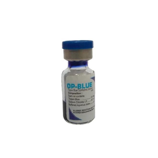
Tired of squinting at tiny letters and pretending you can see them? Modern vision screening devices quietly judge your eyesight in seconds, without the awkward guessing game or eye chart anxiety.
Use automated vision screeners that measure focus, alignment, and eye health quickly and accurately. They reduce human error and improve early detection of problems, as shown in this American Academy of Ophthalmology report.
👁️ Optical Foundations: Refraction, Accommodation, and Light Pathways in Vision Screeners
Modern vision screening devices guide light through the eye and analyze how it bends. By tracking refraction and accommodation, they quickly estimate refractive errors and basic eye health.
These tools use simple optical rules. They project patterns or spots, then record reflected light to map the eye’s focusing power without invasive contact or long exams.
1. Refraction Basics in Automated Screeners
Refraction screening measures how the eye bends incoming light. Devices compare the projected light pattern with the returned image.
- Detects myopia, hyperopia, and astigmatism
- Uses infrared or visible light targets
- Provides fast, objective results
2. Tracking Accommodation and Focus Changes
Accommodation is the eye’s ability to shift focus from far to near. Vision screeners watch these focus shifts in real time.
| Parameter | What It Shows |
|---|---|
| Response speed | How quickly focus changes |
| Range | Near and far focusing limits |
3. Light Pathways and Corneal Reflection
Devices project rings or spots on the cornea and capture their reflections to understand the front surface of the eye.
- Measures corneal curvature
- Helps estimate astigmatism
- Improves alignment and fixation
4. Linking Optics with Retinal Imaging
Some systems combine refraction with retinal imaging. Tools like the Handheld Veterinary Fundus Camera BM-FC-01 and Animal - specific Desktop High - Definition Fundus Camera FS - C1 show how precise optics support clear fundus views.
🔬 Sensor Technologies: From Photorefraction to Wavefront Analysis for Accurate Measurements
Modern sensors read subtle changes in reflected light from the eye. They turn photorefraction and wavefront patterns into detailed, numeric measurements.
These sensors must be stable, fast, and low noise. Their quality strongly affects reliability and repeatability in daily screening.
1. Photorefraction Sensors
Photorefraction uses cameras and infrared light to study light streaks in the pupil.
- Good for quick, binocular screening
- Useful in children and non‑verbal patients
- Works at a short distance
2. Wavefront Sensing Arrays
Wavefront sensors sample many light points across the pupil to map optical errors.
| Type | Key Feature |
|---|---|
| Shack–Hartmann | Lenslet array measures wavefront slope |
| Advanced CMOS | High resolution, low noise imaging |
3. Fundus and Fluorescein‑Based Detection
Some devices use special illumination and dyes to highlight ocular structures. Tools supported by products like Fluorescein Sodium Ophthalmic Strips USP help track tear film and corneal health.
4. Comparative Performance of Key Sensor Types
Different sensor families trade speed, detail, and cost. The chart below compares key attributes for screening tasks.
📊 Data Processing: Algorithms Converting Raw Optical Signals into Clinical Parameters
Software converts raw pixel data and wavefront maps into sphere, cylinder, axis, and risk flags that clinicians can interpret quickly.
Robust algorithms also filter noise, track fixation, and verify consistency between repeated readings.
1. Signal Cleaning and Noise Control
Algorithms remove blinking artifacts and unstable fixation.
- Discard blurred frames
- Average repeated measures
- Flag low‑quality scans
2. Model‑Based Refraction Estimation
Software fits optical models to the eye.
| Output | Use |
|---|---|
| Sphere | Myopia or hyperopia level |
| Cylinder / axis | Astigmatism size and angle |
3. Risk Scoring and Reporting
Devices convert raw values into easy scores. Reports highlight “refer,” “monitor,” or “pass” states, helping teams decide on further exams.
🧒 Pediatric Screening: Noninvasive Principles for Assessing Children’s Visual Function
Pediatric vision screeners use fast, noninvasive optics to check both eyes at once, usually at a short distance with minimal cooperation.
They aim to catch amblyopia risks and refractive errors before school age, when treatment works best.
1. Binocular, Distance‑Based Screening
Devices often work at one meter or more, so children do not feel pressured.
- Simultaneous view of both eyes
- Short capture time
- Kid‑friendly lights and sounds
2. Detecting Amblyopia Risk Factors
Algorithms focus on unequal refraction, strabismus hints, and media opacity.
| Factor | Impact |
|---|---|
| Anisometropia | Unequal focus between eyes |
| High refractive error | Blurry image on the retina |
3. Minimizing Stress and Motion Artifacts
Short tests reduce motion blur and keep children engaged. Clear visual targets encourage stable fixation, improving accuracy.
🏥 Clinical Integration: Workflow, Calibration, and Why Professionals Prefer LumosTail Devices
Vision screeners must fit into clinic workflow, support quick staff training, and maintain calibration across many daily tests.
Reliable hardware plus smart software reduce repeat exams, saving chair time and boosting patient throughput.
1. Streamlined Workflow and EMR Links
Modern devices export results directly to electronic records.
- Automatic patient matching
- PDF and data export
- Clear pass / refer flags
2. Calibration, Maintenance, and Quality Control
Regular calibration ensures stable readings.
| Task | Frequency |
|---|---|
| Self‑check routine | Daily |
| Full calibration | Per service schedule |
3. Why Clinicians Choose Advanced Optical Platforms
Professionals prefer systems that combine strong optics, robust sensors, and easy reporting. This mix delivers trustworthy numbers and clear images that support confident clinical decisions.
Conclusion
Modern vision screening devices blend optics, sensors, and smart algorithms to deliver quick, objective eye measurements. They lower barriers to early detection in both adults and children.
By understanding their working principles, clinics can choose tools that fit their workflow, support accurate referrals, and improve long‑term visual outcomes across diverse patient groups.
Frequently Asked Questions about vision screening devices
1. Are vision screening devices as accurate as full eye exams?
No. They provide fast risk detection, not full diagnosis. Positive or unclear results still need a complete eye exam with an eye care professional.
2. How long does a typical vision screening take?
Most modern devices capture data in a few seconds. Including setup and explanation, a full screening often takes under two minutes per person.
3. Are these devices safe for children and frequent use?
Yes. They use low‑power visible or infrared light that stays within safety limits, making them suitable for repeated use, including in young children.
4. What conditions can modern screeners detect or flag?
They flag refractive errors, amblyopia risk factors, possible strabismus, and sometimes media opacity. They do not replace full retinal or systemic disease evaluation.
5. How often should a clinic calibrate its vision screening devices?
Run built‑in self‑checks daily and follow the manufacturer’s calibration schedule, usually every 6–12 months or after repairs or major software updates.