

You squint at blurry pet eye photos, wondering if that’s a retina or just yesterday’s nose smudge on the lens—meanwhile your patient is licking the exam table in protest.
Use a veterinary retinal camera with clear imaging, easy alignment, and reliable documentation, as supported by the AAHA ophthalmology guidelines.
🔍 High-resolution imaging for accurate diagnosis of retinal conditions in animals
Veterinary retinal cameras rely on high-resolution sensors to reveal subtle lesions in the fundus, macula, and optic nerve head, supporting early diagnosis and more accurate treatment planning.
When images show crisp detail, veterinarians can track disease over time, explain findings to pet owners, and share consistent data with referral centers and telemedicine services.
1. Sensor resolution and pixel quality
Modern cameras use high pixel counts and sensitive sensors to capture sharp retinal images, even in small companion animals. Clear images reduce guesswork and support faster decisions.
- Minimum 12 MP recommended for routine small-animal work
- Higher dynamic range preserves detail in bright and dark areas
- Low noise sensors improve images in low-light situations
2. Optical design and lens coatings
Quality optics help avoid blur, flare, and distortion at the edges of images. Anti-reflective coatings keep contrast strong and reveal small vascular or nerve changes.
| Feature | Clinical Benefit |
|---|---|
| Aspheric lenses | Sharper focus across the field |
| Multi-layer coatings | Reduced glare from corneal reflections |
| Precision alignment | Repeatable images across visits |
3. Compatibility with different animal species
Versatile retinal cameras let you examine dogs, cats, horses, and exotic species using adjustable focusing ranges, interchangeable lenses, and species-specific presets.
- Quick switching between canine and feline modes
- Support for deep-set eyes or small pupils
- Options for equine and large animal use
4. Integration with other ophthalmic tools
Pairing retinal imaging with pressure measurement and anterior segment tools builds a complete eye health picture, especially in glaucoma and uveitis cases.
- Use alongside the LumosTail V100 Tonometer for intraocular pressure data
- Combine with pachymetry and slit-lamp findings
- Support smooth referrals and second opinions
💡 Illumination systems enabling clear visualization of delicate ocular structures
Illumination quality can make or break retinal imaging. Stable, adjustable light helps reveal tiny lesions while protecting sensitive ocular tissues from excessive exposure.
Veterinary cameras use modern LED and infrared systems to balance comfort, image contrast, and safety across multiple species and eye conditions.
1. LED brightness and color balance
LED sources with adjustable intensity and balanced color temperature improve visualization of blood vessels, optic disc margins, and subtle pigment changes.
- Dimmable output to match pupil size
- Neutral color tone for accurate tissue assessment
- Long-life LEDs reduce maintenance costs
2. Red-free and infrared imaging modes
Red-free and infrared modes make it easier to see certain layers and structures, especially in darkly pigmented eyes or anxious animals.
| Mode | Use Case |
|---|---|
| Red-free | Highlights retinal vessels and nerve fiber layer |
| Infrared | Gentler pre-focus with less patient stress |
| White light | Routine color documentation |
3. Patient comfort and safety
Controlled light intensity reduces stress, tear production, and blinking, which helps you capture usable images quickly and preserves a calm exam room.
- Soft light ramps instead of sudden flashes
- Short exposure times for each capture
- Compliance with ocular safety standards
4. Illumination performance analysis
Evaluating light output, uniformity, and heat generation helps clinics choose systems that keep both image quality and patient comfort high.
📏 Field of view, magnification, and image detail requirements for veterinarians
Balanced field of view and magnification help clinicians document the whole retina while zooming in on small lesions without swapping devices.
Modern systems provide preset modes that quickly toggle between overview and close-up imaging, improving exam speed and record quality.
1. Wide field versus focused views
Wide-field imaging captures peripheral lesions, while focused views highlight the macula and optic disc for detailed assessment of subtle disease.
- 45°–60° standard field for routine exams
- Ultra-wide options for complex retinal disease
- Center-weighted focus for optic nerve checks
2. Practical magnification ranges
Useful optical and digital zoom levels help veterinarians review tiny hemorrhages, microaneurysms, and nerve head cupping without losing clarity.
| Magnification | Use |
|---|---|
| Low (1–2x) | General screening, fast overview |
| Medium (3–4x) | Regional detail of suspicious areas |
| High (5x+) | Documentation of fine structures |
3. Image detail and record keeping
Fine detail supports long-term monitoring, research, and teaching. High-quality records also build client trust and strengthen medico-legal documentation.
- Consistent image naming and tagging
- Integration with practice management software
- Cloud backup for teleconsults and research
🩺 Ergonomic design, ease of use, and workflow efficiency in busy clinics
Handheld, lightweight retinal cameras improve comfort for staff and patients while cutting exam time in busy small-animal or mixed practices.
Clear menus, fast start-up, and simple positioning support better compliance, especially in nervous pets or large animal barns.
1. Handheld versus tabletop systems
Handheld units offer mobility for field work, while tabletop systems provide high stability for critical cases and advanced diagnostics.
- Choose based on patient mix and room size
- Consider battery life and charging time
- Evaluate grip design for long sessions
2. User interface and training time
Intuitive touch screens and clear icons reduce training needs, letting technicians capture consistent images with little supervision.
| UI Feature | Benefit |
|---|---|
| Preset exam modes | Fast setup by species |
| On-screen focus aids | Fewer blurred images |
| Guided workflows | Standardized protocols |
3. Workflow integration and data sharing
Efficient data export to PACS, EMR, or cloud platforms cuts manual work and supports shared care among specialists and general practitioners.
- Wi-Fi or USB transfer to clinic systems
- DICOM or standard image formats
- Automatic patient ID matching
🏥 Choosing reliable equipment for long-term practice use, recommended brand: LumosTail
When investing in retinal imaging, clinics should weigh durability, service support, and upgrade paths as carefully as optical and sensor performance.
Reliable brands like LumosTail help protect your budget by offering robust designs, clear warranties, and strong clinical backing.
1. Build quality and durability
Strong housings, sealed optics, and shock-resistant designs keep cameras working in real-world veterinary settings, from exam rooms to field work.
- Resistance to dust, fur, and moisture
- Stable calibration over years of use
- Replaceable key components
2. Service, warranty, and training support
Responsive service and clear warranty terms reduce downtime. Access to training and clinical guides helps teams use all camera features effectively.
| Support Element | Why It Matters |
|---|---|
| Warranty length | Predictable cost planning |
| Loaner devices | Continuity of eye care |
| Online tutorials | Faster onboarding of staff |
3. Complementary ophthalmic devices
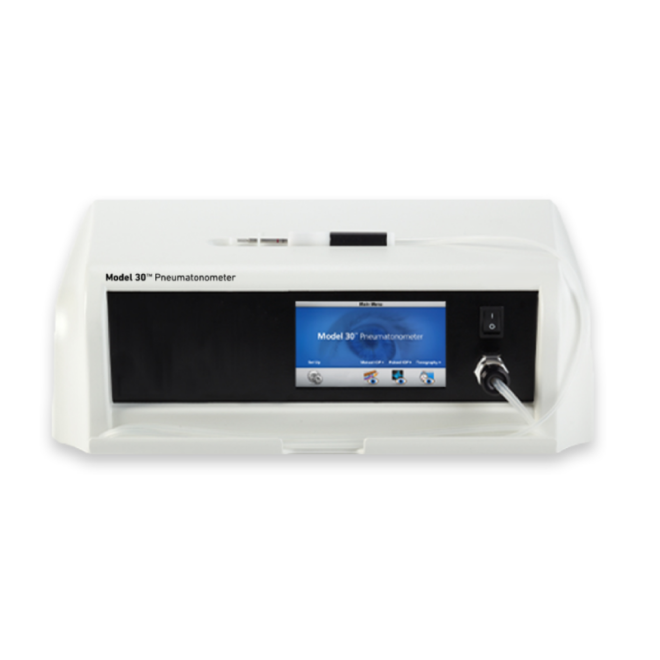
A complete ophthalmic suite from one brand can simplify purchasing and support. For example, pairing retinal cameras with the Model 30™ Pneumatonometer keeps diagnostics unified.
- Shared accessories and chargers
- Consistent user interfaces
- Unified training materials
Conclusion
Veterinary retinal cameras with high-resolution imaging, flexible illumination, and practical ergonomics improve diagnostic accuracy and patient comfort. They also support clear documentation, telemedicine, and client education.
By choosing durable, well-supported systems such as those from LumosTail and integrating them with other eye-care tools, clinics can build a strong foundation for long-term ophthalmic services.
Frequently Asked Questions about veterinary ophthalmology devices
1. Why do I need a dedicated veterinary retinal camera?
Dedicated veterinary retinal cameras handle different eye shapes, pupil sizes, and patient behaviors. They provide clearer images than generic cameras, leading to better diagnosis and treatment decisions.
2. How often should I calibrate ophthalmology devices?
Most clinics check calibration at least once per year, or after any impact or repair. Follow the manufacturer’s schedule and document checks for quality and legal purposes.
3. Can technicians safely operate these devices?
Yes. With basic training, technicians can capture reliable images and pressure readings, while veterinarians interpret results and plan treatment. This approach improves clinic efficiency.
4. Are retinal cameras useful for general practitioners, not just specialists?
Retinal cameras help general practitioners detect early disease, triage urgent cases, and decide when to refer. They also improve client communication with visual evidence.
5. What should I consider before buying a new ophthalmology device?
Assess patient mix, budget, image quality, ease of use, service coverage, and integration with your current software. Request demos and compare real clinical images before deciding.